As demand increases for techniques capable of assessment of the properties of native bone tissue beyond the clinical standard of x-ray densitometry, Raman imaging offers important capabilities for imaging of hydrated tissues. However, its outcomes are currently more varied and less well validated than those of its more mature sibling modality, FTIR imaging. Accordingly, we support our translational FTIR and Raman imaging studies with validation studies of the outcomes of vibrational spectroscopic imaging using chemical standards composed of mixtures of synthetic hydroxyapatite, carbonate-substituted apatite, and collagen.
Key findings: In the validations of FTIR and Raman mineral to matrix ratios—measures of phosphate relative to amide 1—PhD student Erik Taylor showed that the vibrational spectroscopic outcomes were reciprocally related to gold-standard measures of mineral content determined by gravimetric analysis, with native tissue values from humans, sheep, and mice falling on the reciprocal trend established by chemical standards and confirmed by a theoretical analysis. Similarly, in the validations of FTIR and Raman carbonate:phosphate ratios, linear relationships with carbonate content determined by elemental analysis and underlying subbands representing specific carbonate vibrational modes were established. Furthermore, this study identified that FTIR is more sensitive to changes in bone mineral crystal size and composition than Raman spectroscopy. Finally, our multivariate analysis demonstrated that FTIR crystallinity is mostly related to compositional changes in the bone mineral, whereas Raman crystallinity is more influenced by measures of crystal size.
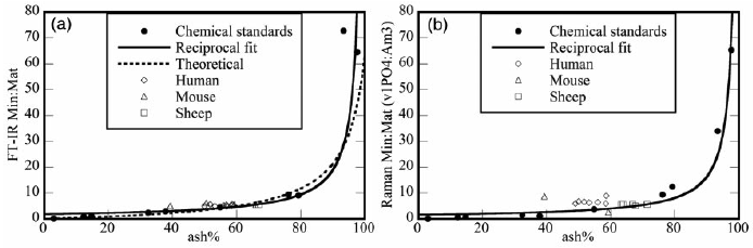
Impact: These validations are the first to relate several Raman mineral:matrix ratio outcomes to ash fraction using gravimetric analysis, the gold standard for analytical characterization of mineral content. Together, these validations now enable biomedical researchers to confidently and efficiently characterize the mineralization and carbonate content of native bone tissue by Raman and FTIR spectroscopy to identify compositional changes related to disease, age, and drug treatment. Furthermore, these validations are an important step in the evolution of Raman spectroscopy as a technique with potential for non-invasive characterization of bone composition.
Future work: We will extend our studies to determine the effects of incident polarization and tissue microstructural orientation on FTIR and Raman crystallinity.

You must be logged in to post a comment.