Individuals with type 2 diabetes mellitus (T2DM) paradoxically have greater fracture risk despite normal or greater BMD relative to non-diabetic individuals, suggesting that impaired glucose metabolism degrades the resistance of bone to fracture. However, the underlying cause of fragility fractures in T2DM is unknown. Over the past several years, we have initiated several new studies of bone tissue material properties in individuals with impaired glucose tolerance and type 2 diabetes
Key findings: In cancellous bone from men with T2DM, bone tissue from the T2DM group had greater modulus, yield stress, and toughness vs. the non diabetic (ND) control group after normalizing for bone volume fraction [Hunt et al. 2019 https://doi.org/10.1002/jbmr.3711]. Specimens from patients with T2DM had greater concentrations of the advanced glycation endproduct (AGE) pentosidine and more mineralized trabeculae compared to non-diabetic controls. The key variables that modulated the energy absorption capability of the tissue were the bone volume fraction, tissue mineral content, and total tissue AGEs. With regression modeling, PhD student Heather Hunt demonstrated that at the 50th percentile of bone volume fraction and mineralization, inclusion of the 90th percentile of AGE content decreased toughness by 36%, suggesting that accumulation of AGEs embrittles bone tissue in men with T2DM.
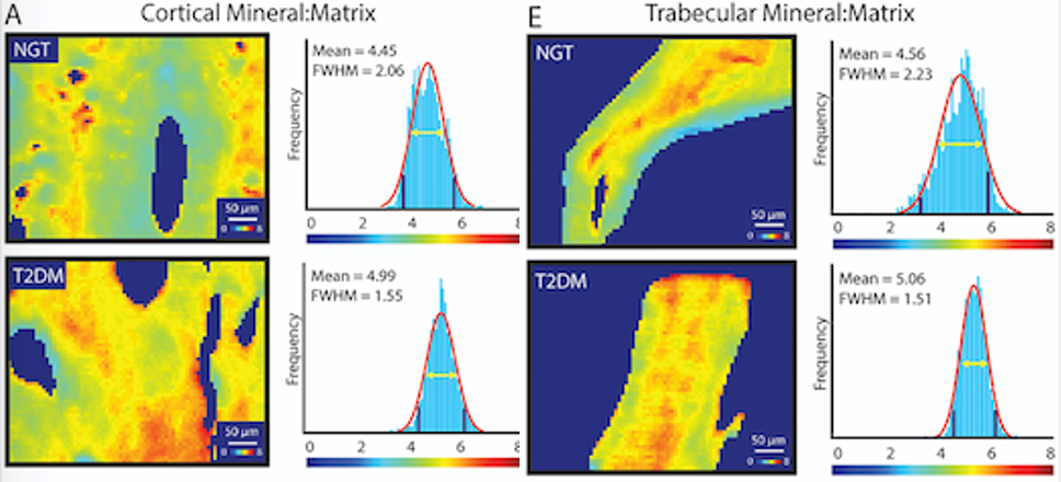
In iliac crest biopsies from postmenopausal women with normal glucose tolerance, impaired glucose tolerance, and overt T2DM, bone tissue from individuals with worsening glycemic derangement had greater cortical and trabecular mineral content and narrower distributions of cortical mineralization [16]. Quantification of bone turnover markers revealed a relatively low turnover state in those with T2DM group compared to those with normal glucose tolerance. Uniquely, our investigation into the mineral properties of bone in T2DM compared to IGT and NGT also supported diminished bone turnover with worsening glycemic control. That is, T2DM was associated with increased mineral content and reduced compositional indicators of new mineral in the bone samples.
In cortical microbeams from the femoral neck, men with T2DM had numerically higher concentration of advanced glycation endproducts across pentosidine HPLC, fluorescence assay, and multiphoton microscopy.
Impact: These data are among the first to characterize the mechanical and biochemical properties of cancellous bone tissue in clinical specimens from patients with type 2 diabetes mellitus. Furthermore, our work supplies the first quantitative data that identifies specific compositional properties contribute to cancellous bone embrittlement in humans with T2DM. This contribution is significant because, in addition to fundamentally advancing the understanding of the pathophysiology of fragility fractures in T2DM, it suggests targets for preventative treatment regimens for this patient population.
Current and future work: Current work includes assessing the effects of T2DM on fatigue failure with bone samples. Future studies include mechanical, biochemical, and/or compositional assessment of bone tissue from several cohorts, including postmenopausal women with varying degrees of glycemic derangement, men and women with T2DM, and women with T1DM, to quantify other novel parameters of bone integrity not captured with traditional screening measures. Additional studies will continue to clarify why BMD fails to accurately predict fracture risk in those with glycemic derangement as well as highlight opportunities for changes in patient screening, fracture prevention, and treatment.
Collaborators:
Deepak Vashishth, PhD: Biomedical Engineering, Rensselaer Polytechnic Institute
Joseph Lane, MD: Orthopedics, Hospital for Special Surgery
Kendall Moseley, Johns Hopkins University School of Medicine
Elaine Yu, Massachusetts General Hospital
Funding: NIH/NIAMS

You must be logged in to post a comment.